Developer Offer
Try ImaginePro API with 50 Free Credits
Build and ship AI-powered visuals with Midjourney, Flux, and more — free credits refresh every month.
AI Now Generates Realistic Retinal Scans
A New Eye for AI GPT-4o's Leap into Medical Imaging
Shortly after OpenAI announced its groundbreaking GPT-4o Image Generation model, researchers immediately began testing its capabilities in a field where AI has traditionally struggled: specialized medical imaging. While Large Language Models (LLMs) have made incredible strides, accurately rendering complex biological structures like ophthalmological images has remained a significant hurdle. A recent report explores whether GPT-4o's enhanced photorealism, as detailed in OpenAI's announcement, could finally bridge this gap.
The First Attempt A Text to Image Test
To begin their investigation, researchers started with a clean slate, disabling ChatGPT's memory feature to ensure no prior conversations could influence the outcome. They gave the model a simple command: “generate a realistic image of a healthy retinal fundus photograph of the posterior pole.”
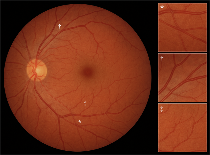
At first glance, the result was impressive and appeared authentic. However, a closer look by a trained eye revealed tell-tale signs of AI fabrication. The retinal background was unnaturally smooth, missing the subtle choroidal vascular patterns seen in real images. Furthermore, the blood vessels displayed atypical characteristics, such as unnatural crossings, an overly bright light reflex, and abrupt changes in their width.
Enhancing Realism with Image to Image Generation
To improve the output, the researchers tried a different approach. They uploaded an authentic fundus photograph, taken from a healthy 49-year-old woman, and prompted GPT-4o to “generate a fundus photograph as similar as possible to this one.” While generative AI cannot create exact copies of uploaded images, it can use them as a powerful reference.
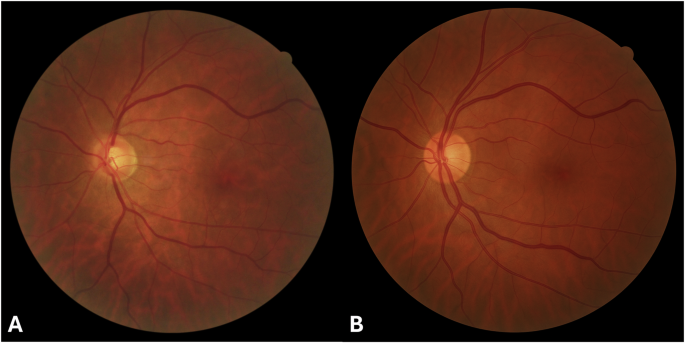
The new image was remarkably more realistic. This time, the AI included the choroidal vasculature, and the retinal vessels appeared anatomically correct, though they still had a slightly pronounced light reflex. The only notable difference was a smaller optic disc cup compared to the original, authentic image. This test demonstrated that with better input, GPT-4o could produce near-authentic results.
The Bigger Picture Training AI for Disease Detection
This capability is more than just a novelty; it has profound implications for the future of medicine. Deep learning models are increasingly used in ophthalmology to detect, classify, and grade retinal diseases, but they require massive datasets of images to be trained effectively. Sourcing these datasets can be challenging due to privacy, cost, and availability.
To solve this, researchers have previously developed generative adversarial networks (GANs) to synthesize high-resolution retinal images. However, developing GANs requires deep technical expertise and significant computational power. The criteria for these synthetic images are strict: they must be realistic enough for specialists to use for diagnosis and be indistinguishable from real photos. The new findings suggest that LLM-based image generation could offer a much faster, cheaper, and more accessible alternative to GANs.
A New Accessible Tool for Medical Research
This study is the first to show that a publicly available LLM like GPT-4o can produce high-resolution, authentic-looking retinal photographs. It marks a significant milestone, potentially democratizing the ability to create synthetic medical data for research and training. While the results are promising, further investigation is needed to confirm if these AI-generated images are robust and varied enough to be included in the training datasets that power the next generation of diagnostic AI.
Compare Plans & Pricing
Find the plan that matches your workload and unlock full access to ImaginePro.
| Plan | Price | Highlights |
|---|---|---|
| Standard | $8 / month |
|
| Premium | $20 / month |
|
Need custom terms? Talk to us to tailor credits, rate limits, or deployment options.
View All Pricing Details