Developer Offer
Try ImaginePro API with 50 Free Credits
Build and ship AI-powered visuals with Midjourney, Flux, and more — free credits refresh every month.
AI Tool Revolutionizes Brain Cancer Diagnosis During Surgery
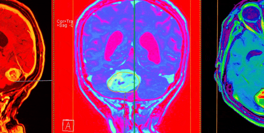 Image: burhan oral gudu/Getty Images
A research team led by Harvard Medical School has created a groundbreaking AI tool that can accurately differentiate between two types of brain cancer that often look identical under a microscope but require vastly different treatments.
Introducing PICTURE A New AI Diagnostic Tool
The tool, named PICTURE (Pathology Image Characterization Tool with Uncertainty-aware Rapid Evaluations), has demonstrated near-perfect accuracy in telling apart glioblastoma and primary central nervous system lymphoma (PCNSL). Glioblastoma is the most common and aggressive type of brain tumor, originating from brain cells. In contrast, PCNSL is a rarer cancer that develops from immune cells. Their microscopic similarities frequently lead to misdiagnosis, which can have severe consequences for patient care.
This innovative work, detailed in an article in Nature Communications, provides a model that is publicly available for other researchers to use and enhance. The tool has also shown to outperform both humans and other AI models in distinguishing between these cancers.
The Challenge of Real-Time Diagnosis
One of the most significant challenges in neuro-oncology is correctly identifying tumors during surgery. An accurate diagnosis while the patient is on the operating table is crucial. It guides the surgeon's decision on whether to remove the cancerous tissue, the standard procedure for glioblastoma, or to leave it and proceed with radiation and chemotherapy, the preferred treatment for PCNSL. A delayed or incorrect diagnosis can result in unnecessary surgery or a delay in starting the correct therapy.
The true value of the PICTURE tool lies in its capacity for real-time deployment during an operation, offering immediate, critical insights to the surgical and pathology teams.
"Our model can minimize errors in diagnosis by distinguishing between tumors with overlapping features and help clinicians determine the best course of treatment based on a tumor’s true identity," stated study senior author Kun-Hsing Yu, an associate professor at Harvard Medical School.
Improving on Current Surgical Procedures
During a typical brain tumor surgery, a tissue sample is removed and rapidly frozen for evaluation under a microscope. While this process provides a quick assessment in about 15 minutes, the freezing can distort cellular features. Based on this initial look, surgeons make the critical call on how to proceed.
A more detailed pathological analysis is conducted over the following days. However, in about one out of every 20 cases, this second, more thorough review changes the initial diagnosis. The PICTURE AI system is designed to fill this gap, dramatically reducing the risk of error when these time-sensitive decisions are made. A key part of its design is an uncertainty feature that flags any tumors the model has not previously encountered, marking them for immediate human review.
Compare Plans & Pricing
Find the plan that matches your workload and unlock full access to ImaginePro.
| Plan | Price | Highlights |
|---|---|---|
| Standard | $8 / month |
|
| Premium | $20 / month |
|
Need custom terms? Talk to us to tailor credits, rate limits, or deployment options.
View All Pricing Details