Developer Offer
Try ImaginePro API with 50 Free Credits
Build and ship AI-powered visuals with Midjourney, Flux, and more — free credits refresh every month.
AI Enhances Medical Scans For Earlier Disease Detection
AI Enhances Medical Scans For Earlier Disease Detection
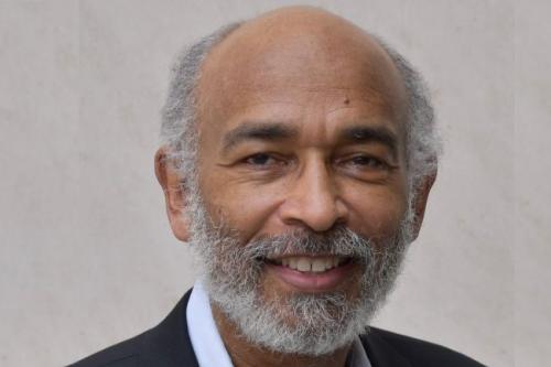
Dr Kofi Deh from Howard Universitys Department of Physics and Astronomy is pioneering research with the Molecular Imaging Laboratory. Their work centers on using Artificial Intelligence AI to improve how we visualize metabolic changes within the body which are crucial indicators for early disease detection.
The Challenge of Early Disease Detection
Changes in metabolism the fundamental chemical processes that keep our cells working can be the very first signs of serious conditions. These include cancer diabetes neurological disorders and many other diseases. Often these metabolic shifts happen long before any structural changes in cells become apparent.
However current medical imaging technologies like Positron Emission Tomography PET scans and Magnetic Resonance Imaging MRI face several hurdles. They often struggle to capture the complete picture of metabolic processes can lack specificity and are frequently expensive. As AI models for image enhancement and analysis continue to advance they are set to play an increasingly vital role. AI can help quickly and accurately spot abnormalities and metabolic shifts paving the way for faster more precise diagnoses and subsequent treatments.
AI to the Rescue Super Resolving Medical Scans
Dr Dehs research focuses on significantly improving the visual quality of medical scans. He employs an artificial neural network to super resolve these images. This advanced technique requires two key components naturally high quality images to serve as a benchmark and detailed data on the physical processes being imaged.
The Science Behind AI Enhanced Imaging
Dr Deh explained that acquiring the higher resolution images needed for training the neural network requires resorting to physics informed methods where one actually models the processes being targeted for super resolution. This means the AI is not just guessing it is learning from the underlying physics of what it is trying to see.
A Practical Example Imaging Brain Activity
Dr Deh provided an illustrative example involving the measurement of diffusion in human brains. Diffusion is the gradual spread of molecules from areas of high concentration to areas of low concentration.
He clarified that one cannot measure diffusion at each point in the brain. However pressure can be measured at several points in the brain or at several arteries. Doing so allows for the creation of equations that model blood flow.
These mathematical models of blood flow when combined with anatomical images are then fed into the neural network. This ensures that the image enhancement process is not only visually sharper but also accurately reflects the biological reality.
The Future of Medical Diagnosis
The integration of sophisticated AI techniques like those being developed by Dr Deh holds immense promise for the future of medical diagnostics. By providing clearer more detailed and more accurate images of metabolic functions clinicians can gain earlier insights into disease development. This can lead to more timely interventions personalized treatment plans and ultimately improved patient outcomes. The ongoing refinement of these AI driven imaging methods signals a significant step forward in our ability to combat complex diseases.
Further Insights and Media Information
To explore more research and news from Howard University:

If you are a member of the media Howard Universitys public relations team can connect you with faculty experts and provide information about university news and events. You can Submit a Media Inquiry.
Compare Plans & Pricing
Find the plan that matches your workload and unlock full access to ImaginePro.
| Plan | Price | Highlights |
|---|---|---|
| Standard | $8 / month |
|
| Premium | $20 / month |
|
Need custom terms? Talk to us to tailor credits, rate limits, or deployment options.
View All Pricing Details